A concept for a novel X-ray microscope promises three-dimensional images of delicate objects like biological cells using a thousand times less damaging radiation than conventional methods. The novel microscope would allow to image whole cells at high resolution in their native environment, without freezing, cutting or staining them. Scientists Dr. Pablo Villanueva-Perez, Dr. Saša Bajt and Prof. Henry Chapman from the Center for Free-Electron laser Science (CFEL) present their concept in the journal Optica. The simulation study yields a bright perspective for the planned upgrade of DESY’s storage ring PETRA III to a next generation X-ray source, PETRA IV.
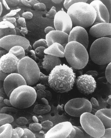
Human blood cells under the scanning electron microscope. Credit: National Cancer Institute [Source]
Imaging the structures of biological cells on the nanometre scale usually requires X-rays, as their short wavelengths allow to resolve the fine details. “However, X-rays also deposit energy that quickly damages biological samples,” says DESY scientist Villanueva-Perez. How fast radiation damage occurs depends on the characteristics of the object under study and on the energy of the X-rays used, but in practice is the limiting factor for resolution and sensitivity of today’s X-ray imaging techniques.
X-ray images can be formed by a variety of means. The familiar radiographs of teeth or broken bones rely on absorption — the dense bone leaves a dark shadow in the image where X-ray photons are absorbed. An X-ray microscope built for imaging cells usually depends upon elastic scattering of the X-rays in the sample to achieve images of much higher resolution. This is similar to how images are formed in an optical microscope. Although elastic X-ray scattering transfers no energy, in all X-ray microscopes built to date, such scattering processes happen much less frequently than actual absorption. “In reality, scattering cannot occur without a fraction of the photon’s energy being deposited in the sample, producing radiation damage,” says Villanueva-Perez.
Objects become much less absorbing the more energetic the X-ray photons. However, such high energies were not considered useful for high-resolution microscopy since the elastic scattering also decreases and another form of scattering becomes predominant. In this inelastic process, also known as Compton scattering, the X-ray loses some of its energy to the object as it ricochets off an atom and in the process changes wavelength. This usually produces unwanted featureless background or fog in the image, deteriorating the quality of both the image and the sample.
The insight of the team was that at very high X-ray photon energies of 64 kiloelectronvolts (keV) there are many more Compton scattering events for a given amount of energy deposited into the cell than for elastic scattering at the conventional lower photon energies exploited by current techniques. A detailed image can then be built up by rastering a focused X-ray spot across the cell and mapping out the total scattering detected at each location. Surprisingly, analysis showed that the dose could be reduced by a factor of 1000 for a given resolution. “No one really thought of trying biological microscopy at such high energies,” explains CUI member Chapman (Universität Hamburg, DESY). “Bright enough X-ray sources didn’t exist, there was no way to focus the beam, and there were no detectors.”
The team has found solutions to these challenges. Bajt’s team just recently developed an innovative lens from an artificial multilayer “metamaterial” that delivers the smallest X-ray focus yet achieved. “The efficiency of our multilayer lenses actually gets much better with increasing energy, and they make even smaller spots,“ says DESY scientist Bajt. “So they are ideally suited to building our microscope.”
The X-ray source PETRA IV, currently in the planning phase, will deliver beams of much higher X-ray brightness at the required high photon energies than possible today. This still leaves the detector. “The ideal detector should surround the sample, to collect all scattered photons in all directions,” explains Villanueva-Perez. This can be built using today’s technology. Once realised, these ingredients will enable scientists to scan whole cells and organelles at a few nanometres resolution in all three dimensions, in their natural state – fulfilling a widespread wish of biologists. Until then, the scientists plan to test their novel concept with biological samples at today’s best X-ray sources like PETRA III with conventional detectors. Text: DESY, ed.
Citation:
Pablo Villanueva-Perez, Saša Bajt, and Henry N. Chapman
Dose efficient Compton X-ray microscopy
„Optica“, 2018
DOI: 10.1364/OPTICA.5.000450